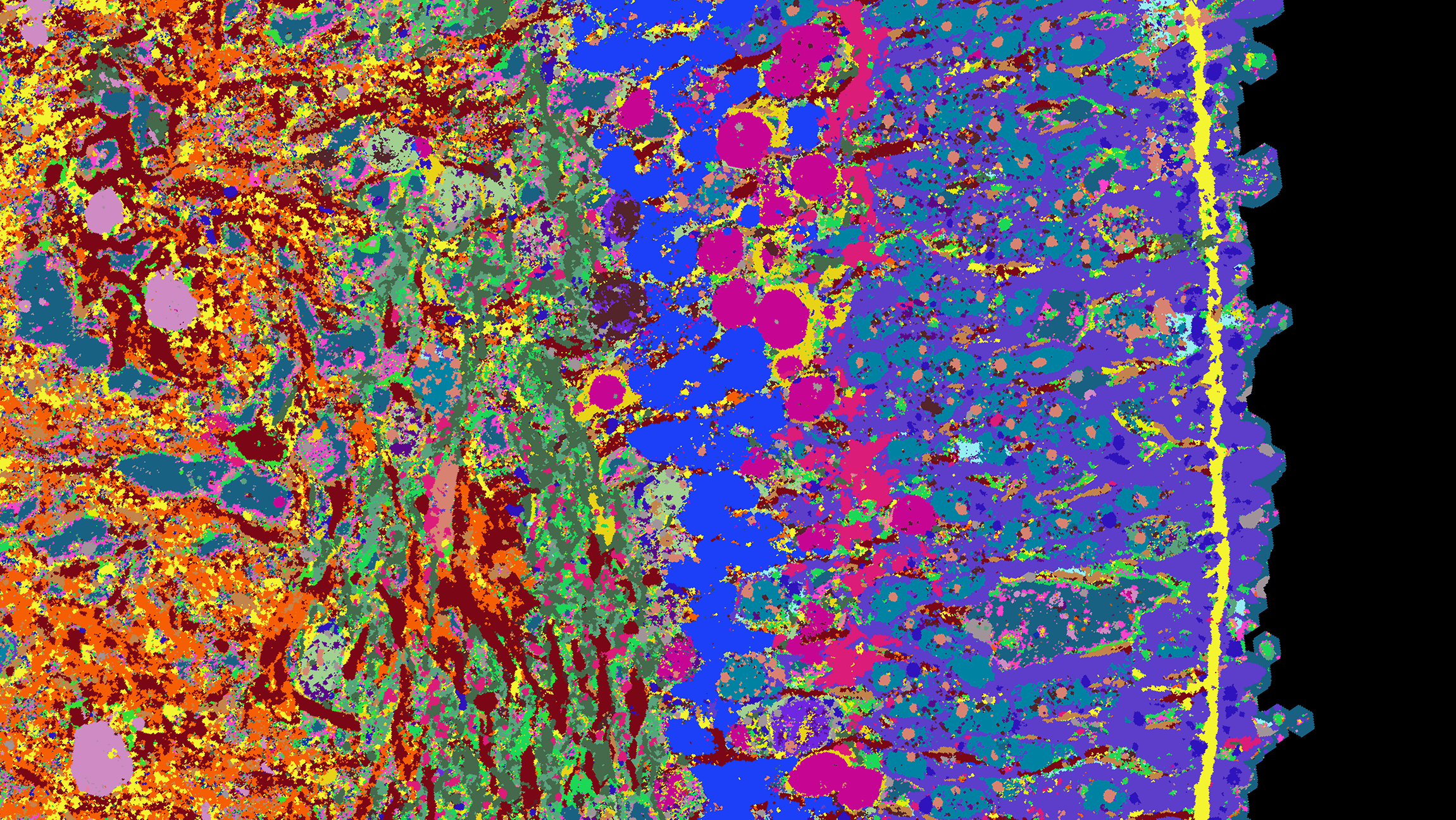
Detailed image of the human retina
Researchers from Basel and Zurich are creating a high-resolution atlas that depicts the development of the human retina. One technique they use is a new method that allows them to visualise more than 50 proteins simultaneously.
Abstract
- A new atlas illustrates how the human retina is developing.
- To create it, researchers mapped three-dimensional tissue structures called organoids that they had cultivated in their lab.
- The atlas could support research to halt the progression of a degenerative eye disease that can cause people lose their sight.
What cell types are found in which human tissue, and where? Which genes are active in the individual cells, and which proteins are found there? Answers to these questions and more are to be provided by a specialised atlas – in particular how the different tissues form during embryonic development and what causes diseases. In creating this atlas, researchers aim to map not only tissue directly isolated from humans, but also structures called organoids. These are three-dimensional clumps of tissue that are cultivated in the laboratory and develop in a way similar to human organs, but on a small scale.
“The advantage of organoids is that we can intervene in their development and test active substances on them, which allows us to learn more about healthy tissue as well as diseases,” explains Barbara Treutlein, Professor of Quantitative Developmental Biology at the Department of Biosystems Science and Engineering at ETH Zurich in Basel.
To help produce such an atlas, Treutlein, together with researchers from the Universities of Zurich and Basel, has now developed an approach to gather and compile a great deal of information about organoids and their development. The research team applied this approach to the organoids of the human retina, which they derived from stem cells.
Read on > full-length ETH News.

“The advantage of organoids is that we can intervene in their development and test active substances on them, which allows us to learn more about healthy tissue as well as diseases.”Barbara Treutlein, Quantitative Developmental Biology lab, D-BSSE, ETH Zurich
Find the full-length ETH News (in English and German).
Learn about the Quantitative Developmental Biology lab led by Barbara Treutlein.