Organoid Atlas: Understanding tissue development and degeneration
The eye-catching image in the current issue of GLOBE was taken by the group of Quantitative Developmental Biology group of Barbara Treutlein. It shows a thin section of retinal tissue, stained with fluorescent dyes and imaged using fluorescent microscopy. This process was developed to image 3D issue structures cultivated in the lab. A comprehensive atlas now combines all images from organoids to improve our understanding of tissue development and degeneration.
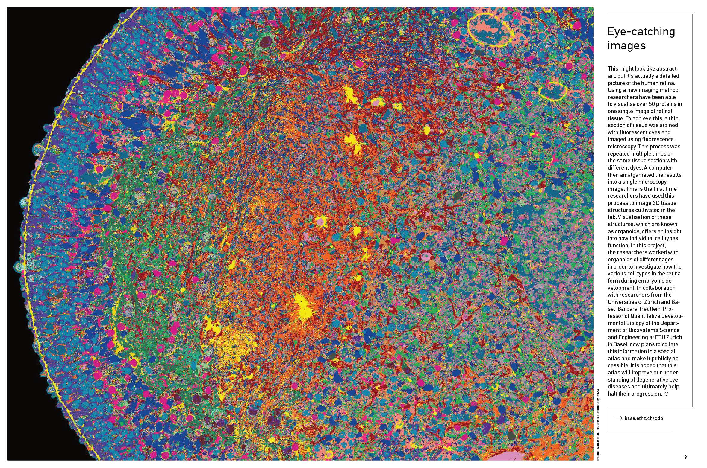
This image was published by Download ETH Zurich's GLOBE magazine 3/2023 (PDF, 4.6 MB) (p. 8-9).
Learn about research in the Quantitative Developmental Biology lab led by Barbara Treutlein.