Electrophysiology & Neuroscience
external page Electrophysiology is the study of the electrical properties of biological cells and tissues. It involves measurements of voltage changes or electric currents on a wide variety of scales from single ion channels to whole organs like the heart. In neuroscience, it includes measurements of the electrical activity of neurons and, particularly, action potential activity.
external page Neuroscience is the scientific study of molecular, cellular, developmental, structural, functional, evolutionary, computational, and medical aspects of the nervous system. It is an interdisciplinary science that includes aspects of biology, chemistry, computer science, engineering, mathematics and medicine.
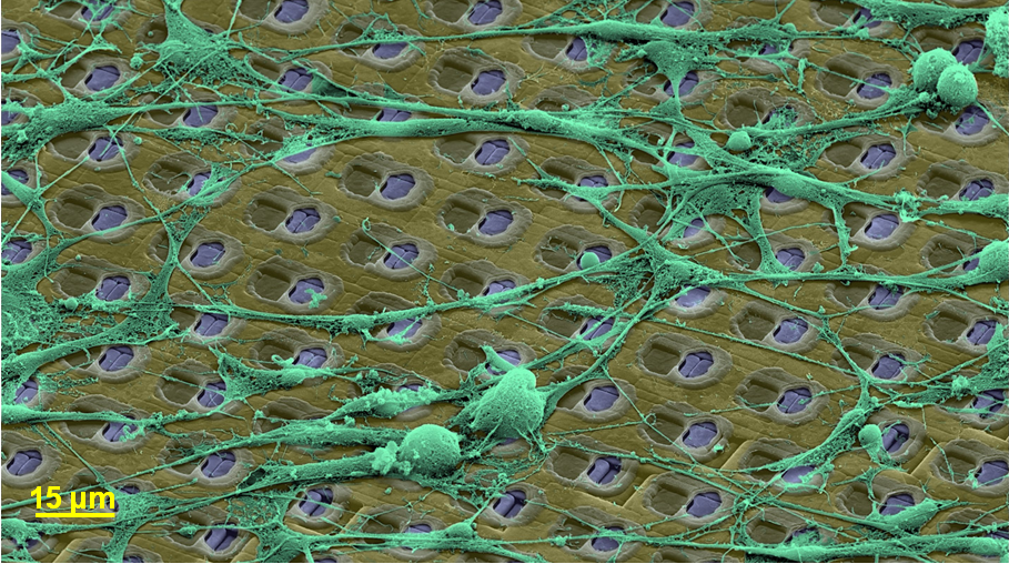
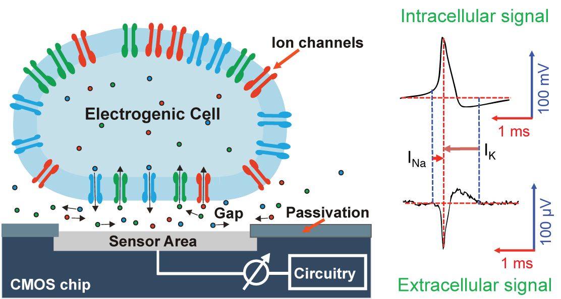
We pursue an extracellular, bioelectronic approach to electrophysiology, which relies on bringing electrogenic cells (cells that produce electrical activity) and tissues in direct contact with state-of-the-art integrated electronic systems.
The cell preparations (dissociated cells, tissue slices) are placed directly atop fully processed microelectronics chips carrying thousands of electrodes and featuring CMOS circuitry. The bio-electronic interface consists prevailingly of noble-metal electrodes.
Prospective fields of applications of our technology include, besides fundamental neuroscience research, pharmascreening, the investigation of mechanisms of neurodegenerative diseases, or the development of aural and visual prostheses.
(1) Human-derived Neurons and Organoids
We apply the high-resolution microelectrode technology to study effects of developmental or neurodegenerative diseases on neuronal functioning in collaboration with academic and industrial partners. We use mostly human-derived neuronal preparations. i.e., neurons generated from human induced puripotent stem cells (hiPSCs). Besides dissociated neurons, we use organoids as well acute and organotypic slice preparations of rodent origin.
More information
(2) Retinal Investigations
The retina is an almost 2-dimensional neuronal system, for which high-density microelectrode arrays are ideally suited to record from the retinal ganglion layer. By applying light stimuli, we try to find defined retinal ganglion cell types, to characterize their behavior and to decipher features of retinal coding of visual inputs. Moreover, we participate in efforts of human visual restoration.
More information
(3) Combination of Techniques
Besides the extensive use of our high-density microelectrode arrays (HD-MEAs) for extracellular electrophysiology across scales - from subcellular compartments through individual neurons to entire networks - we also devlop combined techniques, which enable HD-MEA recordings with simultaneous confocal microscopy, patch clamp or atomic-force measurements on the same chip. Moreover, we develop HD-MEA technology extensions like the use of wire bundles or printed 3D electrodes to record in vivo.
More information
(4) Neuronal Signal Analysis
The multitude of tightly spaced electrodes produces large data sets, where every neuron is recorded from by many electrodes, and where every electrode records signals from many neurons. This poses challenges to spike sorting and signal processing. We develop tools and methods to extract meaningful information from the large datasets and use the information for detailed modeling of neuronal behavior.
More information