Retinal Investigations
The vertebrate external page retina is a light-sensitive layered tissue, lining the inner surface of the eye. The optics of the eye create an image of the visual world on the retina (through the cornea and lens), which serves much the same function as the film in a camera. Light striking the retina initiates a cascade of chemical and electrical events that ultimately trigger nerve impulses. These are sent to various visual centers of the brain through fibers of the optic nerve.
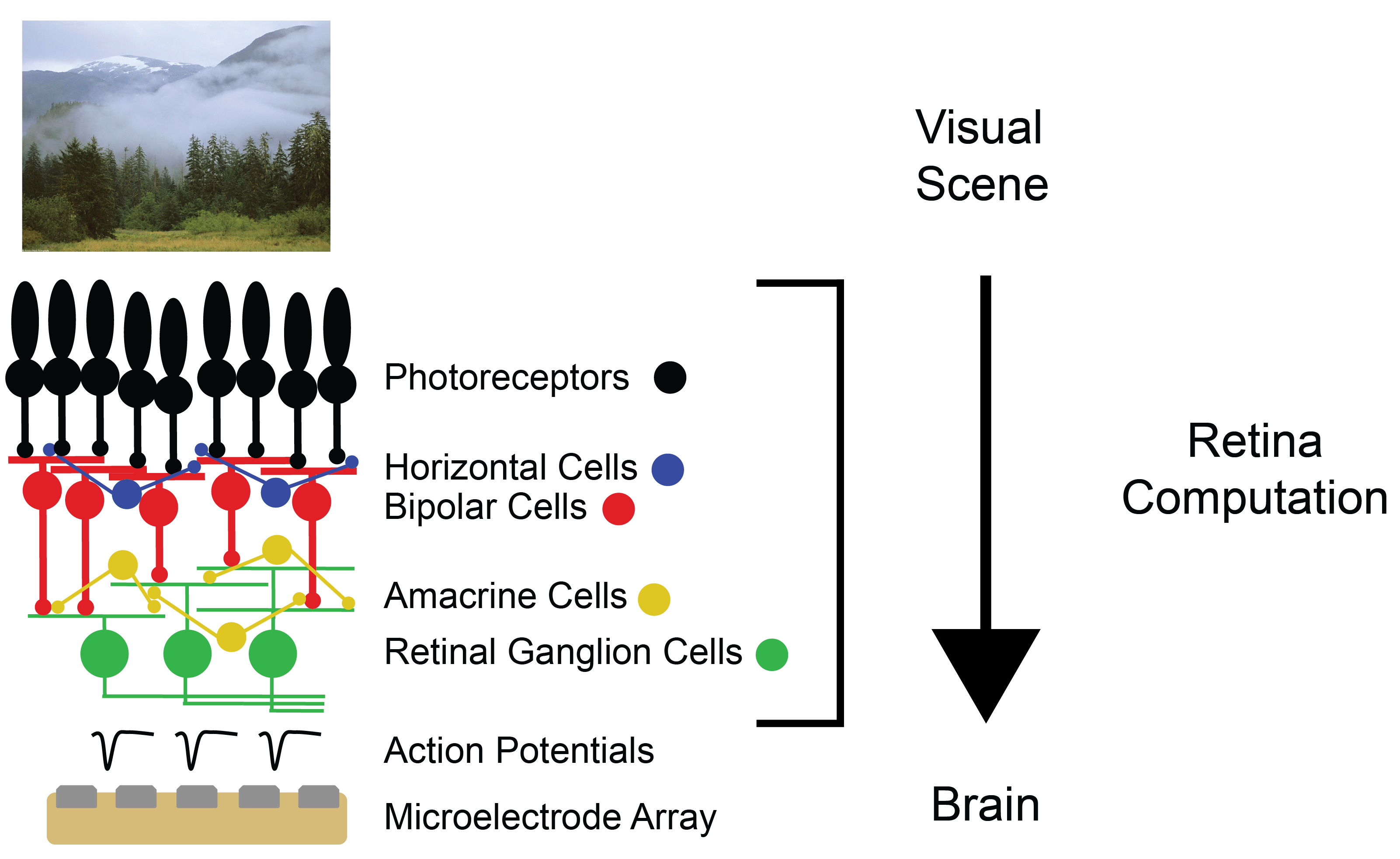
The retina converts visual input into electrical cell activity (action potentials), and retinal ganglion cells (RGCs), which are organized in a two-dimensional arrangement, form the output layer of the retina. There are approximately 20 RGC types that convey information about a visual scene to the brain, while information and signal processing is performed already in the neuronal circuits in the retina.
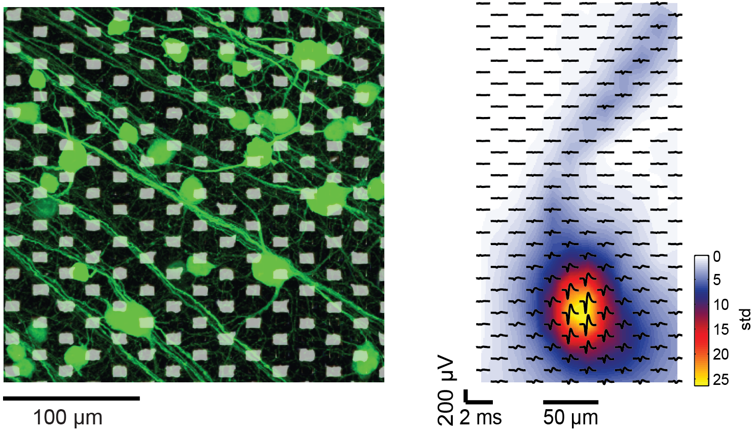
We are interested to contribute to deciphering the coding of visual input in the retina and to find out how the brain interprets retinal signals. Therefore, we apply high-resolution microelectrode arrays (MEA) technology to trace electrical external page action potentials of multiple RGCs at subcellular resolution. The technology enables us to precisely identify the different RGC types according to their behavior upon light stimulation and to simultaneously record the activity of multiple ganglion cells of the same or different types.
Collaborations
Friedrich-Miescher Institute, Basel, Switzerland; University of Tübingen, Germany; Ecole Nationale Supérieure, Paris, France.
Recent publications
R. Diggelmann, M. Fiscella, A. Hierlemann, F. Franke, "Automatic Spike Sorting Algorithm for High-Density Microelectrode Arrays", Journal of Neurophysiology 2018, 120 (6), pp. 3155–3171 (DOI: 10.1152/jn.00803.2017). external page Online
A. Drinnenberg, F. Franke, R. K. Morikawa, J. Jüttner, D. Hillier, P. Hantz, A. Hierlemann, R. A. da Silveira, B. Roska, "How diverse retinal functions arise from feedback at the first visual synapse", Neuron 2018, Vol. 99 (1), pp. 117-134.e11 (DOI: 10.1016/j.neuron.2018.06.001). external page Online / external page Preview
K. Yonehara, M. Fiscella, A. Drinnenberg, F. Esposti, S. Trenholm, J. Krol, F. Franke, B. Gross-Scherf, A. Kusnyerik, J. Müller, A. Szabo, J. Jüttner, F. Cordoba, A. P. Reddy, J. Németh, Z. Nagy, F. Munier, A. Hierlemann, B. Roska, "Congenital nystagmus gene FRMD7 is necessary for establishing a neuronal circuit asymmetry for direction selectivity", Neuron, 2016, 89(1), pp. 177-193 (DOI: 10.1016/j.neuron.2015.11.032). external page Online / external page Video abstract
F. Franke, M. Fiscella, M. Sevelev, B. Roska, A. Hierlemann, R. Azeredo da Silveira, "Structures of neural correlation and how they favor coding", Neuron, 2016, 89(2), pp. 409-422 (DOI: 10.1016/j.neuron.2015.12.037). external page Online
M. Fiscella, F. Franke, K. Farrow, J. Müller, B. Roska, R. Azeredo da Silveira, A. Hierlemann, "Visual Coding with a Population of Direction-Selective Neurons", Journal of Neurophysiology, 2015 (DOI: 10.1152/jn.00919.2014). external page Online