Resistance and Impedance Measurements
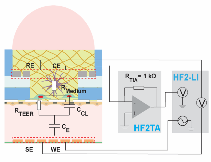
Resistance and impedance measurements constitute a label-free and nioninvasive method to characterize cells and tissues. Transepithelial/transendothelial electrical resistance (external page TEER) measurements are used to monitor the tightness of membranes or in-vitro models of physiological barriers, such as the lung or blood-brain barrier (BBB).
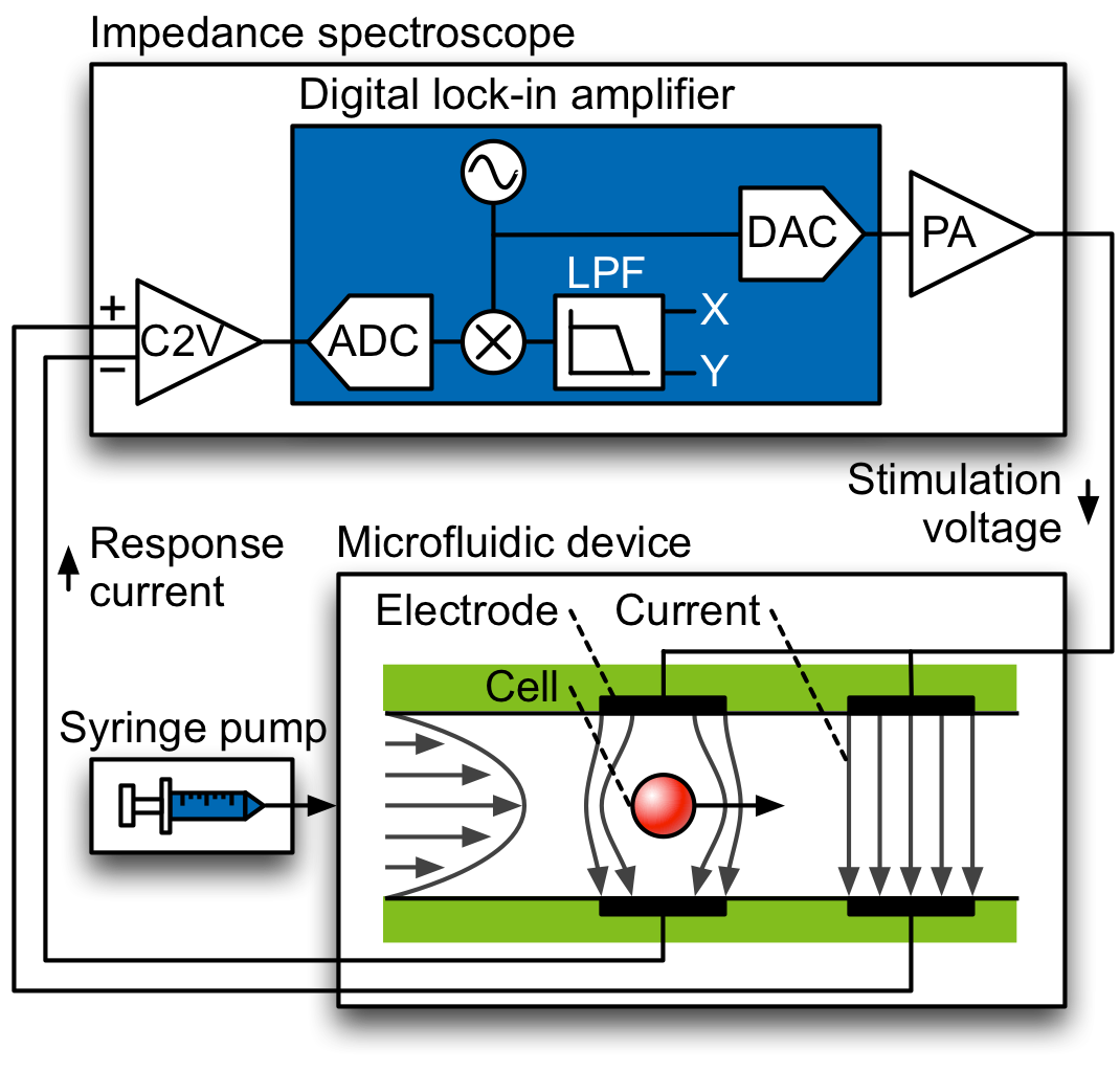
Electrical impedance spectroscopy (external page EIS) is a technique for measuring the dielectric properties of a material of interest in dependence of the applied frequency. The method is used in the biological domain to characterize cells and tissues. The dielectric properties reveal information about cell size, membrane resistance, membrane capacitance and cytoplasmic conductivity. To conduct measurements, cells or particles are typically dispersed in an electrolyte, such as phosphate-buffered saline (PBS), and forced through a microfluidic channel with two pairs of planar electrodes patterned at the top and bottom. An AC voltage is applied to the electrodes, and the current change upon passage of a particle is measured differentially.
In collaboration with external page Zurich Instruments AG, we have developed impedance cytometers capable of measuring the dielectric properties of single cells at frequencies of up to 600 MHz. The increased frequency range can be used to characterizing subcellular features, such as vacuoles and cell nuclei, in addition to the properties detectable at lower frequencies.
Impedance spectroscopy can be combined with other electrical functionalities in microfluidic chips, such as application of strong electric fields to induce pore formation in the cell membrane. These can be used to extract cellular contents for imaging or spectrometry or to introduce external material into the cell for labeling or external page transfection.
Relevant publications
R. Bounik, A. Landolt, J. Lee, V. Viswam, F. Cardes, M. Modena, and A. Hierlemann, "Seamless integration of CMOS microsensors into open microfluidic systems", Lab on a Chip 2025, 25, pp. 2205 - 2221 (DOI: 10.1039/D4LC01000K). external page Online
W. Wei, F. Cardes, A. Hierlemann, M. Modena, "3D in vitro blood-brain-barrier model for investigating barrier insults", Advanced Science 2023, Article 2205752 (DOI: 10.1002/advs.202205752). external page Online
R. Bounik, F. Cardes, H. Ulusan, M. Modena, A. Hierlemann, "Impedance imaging of cells and tissues: design and applications", BME Frontiers 2022, Article 9857485 (DOI: 10.34133/2022/9857485). external page Online
F. Gökçe, P. S. Ravaynia, M. M. Modena, A. Hierlemann, "What is the future of electrical impedance spectroscopy in flow cytometry?", Biomicrofluidics 2021, 15, 061302 (DOI: 10.1063/5.0073457). external page Online
P. S. Ravaynia, F. C. Lombardo, S. Biendl, M. A. Dupuch, J. Keiser, A. Hierlemann, M. M. Modena, "Parallelized impedance‐based platform for continuous dose‐response characterization of antischistosomal drugs", Advanced Biosystems 2020, Article 1900304 (DOI: 10.1002/adbi.201900304). external page Online