HD-MEAs for performing functional imaging of brain organoids
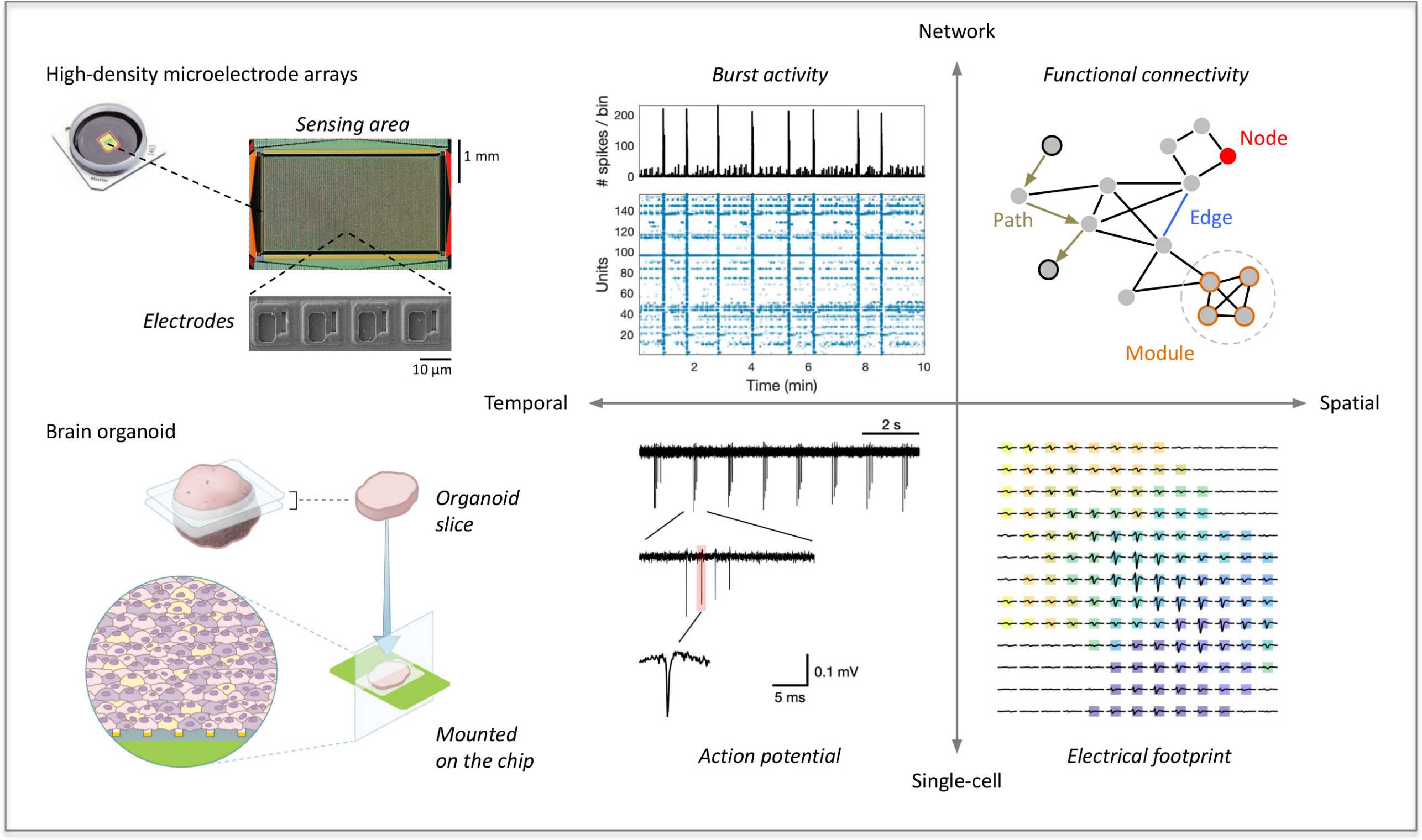
New BEL paper titled "Functional imaging of brain organoids using high-density microelectrode arrays" describes use of planar microelectrode arrays for characterizing human pluripotent-stem-cell derived 3D brain organoids.

Human cerebral organoids (hCOs) recapitulate fundamental milestones of early brain development, but many important questions regarding their functionality and electrophysiological properties remain open. High-density microelectrode arrays (HD-MEAs) can be used to perform functional studies of neuronal networks at cellular and network scales. In the most recent paper in the Impact section of the MRS Bulletin, Manuel Schröter et al. describe the use HD-MEAs to derive large-scale electrophysiological recordings from sliced hCOs (M. Schröter, C. Wang, M. Terrigno, P. Hornauer, Z. Huang, R. Jagasia, A. Hierlemann, "Functional imaging of brain organoids using high-density microelectrode arrays", MRS Bulletin 2022, Vol. 47). Electrical activity of hCO slices was recorded over several weeks, spike-sorted and subsequently studied across scales. Single neurons were tracked over several days on the HD-MEA, and axonal action potential velocities were determined. The developed methodology will contribute to a better understanding of neuronal networks in brain organoids and provide new means for their functional characterization. The work was a collaboration with F. Hoffmann-La Roche Ltd., the Roche Innovation Center in Basel.
external page MRS Bulletin Impact is a monthly published premium selection of the MRS Bulletin of new, original, and important research that demonstrates a significant impact or advance in scientific understanding of exceptional interest to the materials research community.
