Point-scanning Confocal Microscopes
The Single Cell Facility is equipped with multiple point-scanning Confocal Micoscopes. An overview is given below.
For detailed descriptions of the individual workstations (all specifications, filter settings, etc.) and interactive spectra viewers, see our internal Wiki (D-BSSE login required).
Nikon Confocal microscopes
The Nikon confocal microscopes are fully motorized confocal systems that excel through their modularity. Optimized for the higher resolution 3D-imaging of both live and fixed samples, they offer a flexible choice for variety of experiments, including z-stacks for 3D reconstruction, 3D time-lapse imaging (XYZT), multipoint time-lapse imaging, colocalization studies, large field imaging (stitching), spectral analysis (AXR), FRAP, FLIP, FRET, and photoactivation. Each setup is equipped with an environmental control chamber that allows for live cell support (temperature, CO2, %RH). In addition, each microscope can be custom-scripted to perform more complex and/or ‘smart’ imaging routines through the unique JOBS software module.
Nikon A1 HD25 Confocal (Nikon WF6 + A1)
The Nikon A1 HD25 confocal microscope is a younger, ‘simpler’ version of the same scanner series, which makes use of the newer generation Ti2 design to offer a 39% larger field of view. It is mounted on a combined setup, allowing full functionality of both wide field and confocal imaging options. Moreover, both modalities are fully integrated and combinable in advanced imaging routines using the JOBS software module.
- Microscope base: Nikon Eclipse Ti2-E (inverted)
- Confocal scan head: Nikon A1 HD25
- Scanner: galvano scanner 1Hz-1400Hz
- Detection mode: Standard filter-based detector
- Detectors: 3x PMT (1 for transmission) + 2x GaAsP (standard) + 32 PMT-array (spectral)
- WF light path: right port
- WF Light engine: Lumencor Spectra X (387/438/466/500/531/632 nm)
- WF Camera: Andor Sona 4.2B-11
- Special: combination with wide field imaging, large FOV scanning, deep-learning based Poisson shot noise removal (Denoise.ai)
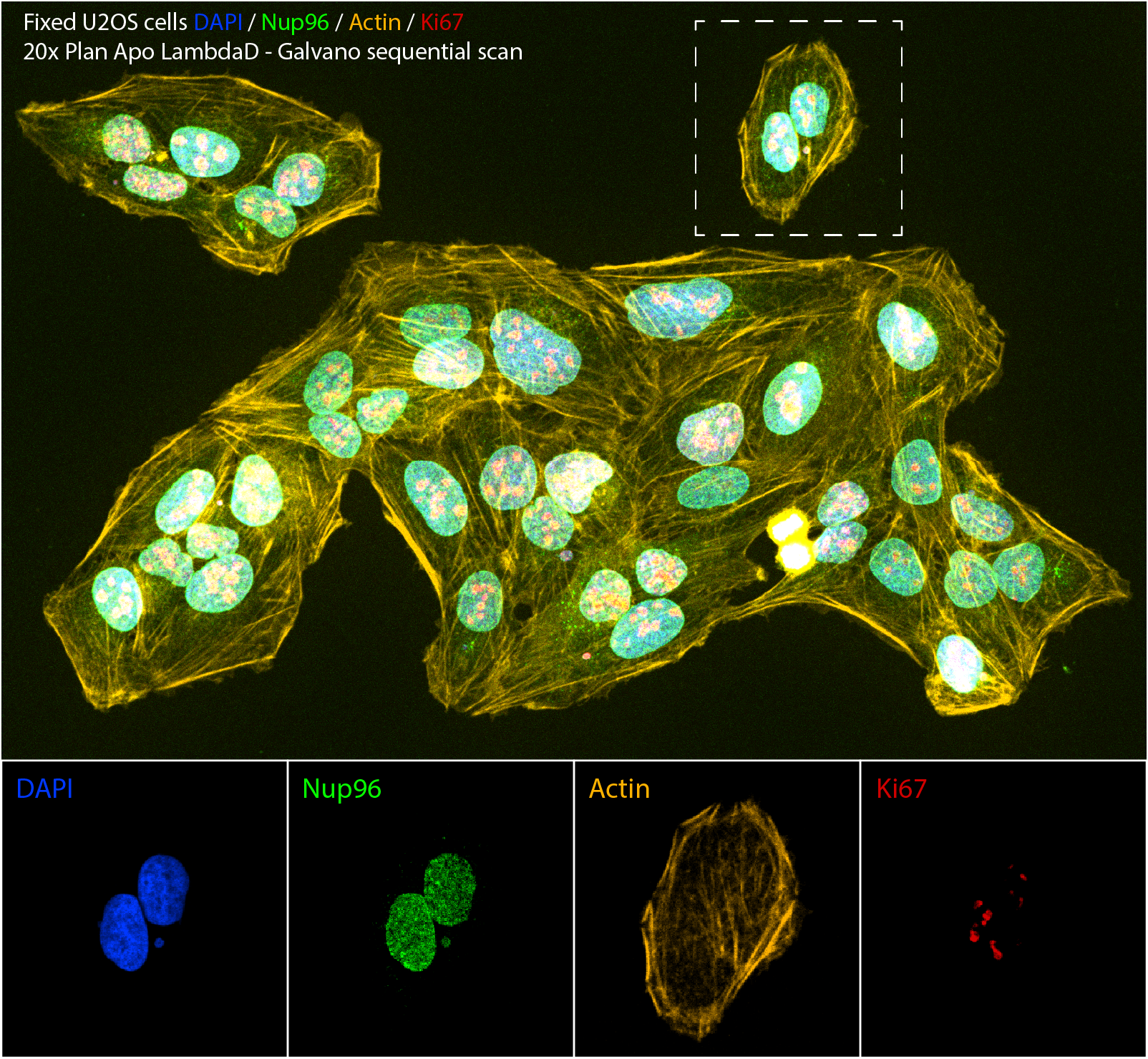
Nikon AXR Confocal (Nikon AXR)
The Nikon AXR confocal microscope is a 4-laser system equipped with a hybrid resonant + galvano scanner, which makes use of the newer generation Ti2 design to offer a 25mm diagonal field of view. The hybrid scanner design allows for fast imaging (2k x 2k pixels with the resonant scanner) as well as high sampling (8k x 8k) and e.g. localized photoactivation using the galvano scanner. To improve sensitivity and flexibility, the system features 2 GaAsP PMT detectors with variable spectral filters (max. range 400-750nm), in addition to two standard multi-alkali PMTs in the blue and far red channels. Various acquisition setups are possible due to the availability of the variable DUX-VB detector unit, including (sequential) spectral imaging at a minimum bandwidth of 5 nm. Equipped with the dedicated Plan Apo LambdaD objective series to make maximum use of the large FOV.
- Microscope base: Nikon Eclipse Ti2-E (inverted)
- Confocal scan head: Nikon AXR
- Scanner: galvano scanner (30 fps max. at 512 x 512 pixels) + resonant scanner (30 fps max. at 512 x 512 pixels)
- Detection modes: Standard filter-based detection on 2 PMTs, variable spectral filtering on 2 GaAsP detectors
- Detectors: 3x PMT (1 for transmission) + 2x GaAsP (default)
- Piezo z-stage: Mad City Labs Nano-Drive (200 µm)
- WF light path: right port
- WF excitation: Nikon D-LEDI
- WF filter cubes: DAPI, GFP, Cy3, Cy5
- WF Camera: Hamamatsu Orca Flash v2
- Special: 2k resonant scanner, dual variable filter GaAsP detectors, large FOV (25mm) compatibility, combination with wide field imaging, integrated AI functions (AutoSignal.ai, Denoise.ai, etc.)
Leica TCS Confocal microscopes
Leica TCS confocal microscopes are filter free microscopes. The acousto-optical beam splitter (AOBS) that separates excitation and emission light, together with the prism-based spectral dispersion of the emission light, gives a high freedom in the dye selection. Each includes at least 9 excitation laser lines and 5 detectors, with the possibility to precisely define the emission light band to be directed to the detectors.
All our Leica confocals are equipped with 3 standard PMTs and 2 very-high-sensitivity HyD detectors. The HyD‘s very good dark noise behavior, combined with its higher quantum efficiency, makes them ideally suited for high-speed imaging using the resonant scanner, minimizing light exposure of the specimen.
The microscopes are also equipped with an environmental control chamber that allows short and long term live cell imaging (Temperature, %CO2, %RH).
Leica SP8 Confocal (Leica SP8)
Our SP8 confocal features the LAS X Navigator software module, generating live overviews to identify regions of interest for multiparametric acquisition and large-scale (up to several cm) tile acquisitions in a very user-friendly and robust way. The integration with the hardware-based autofocus system facilitates keeping the sample in focus over time and space (distance).
- Microscope base: Leica DMI 8
- Leica TCS Tandem scanner: standard scanner 10Hz-1400Hz + resonant scanner 8000Hz
- 2 HyD detectors, 3 PMT Detector + Transmitted light
- Available lasers: 405, 442, 458, 476, 488, 496, 514, 561, 594, 633 nm
- Objectives (are subject to change): 10x, 20x (dry and multi immersion oil/glycerol), 40x, 63x (water and oil immersion)
- LAS X Navigator
- Hardware autofocus system
Leica SP8-FALCON Confocal (Leica SP8-FALCON)
The Leica SP8-FALCON (FAst Lifetime CONtrast), has all the features of our other SP8, including the LAS X Navigator module and the Hardware autofocus system. In addition, thanks to the integrated hard- and software module for Fluorescence Lifetime Imaging (FLIM), it offers a new dimension of contrast to the classical fluorescence confocal acquisition. Now it is possible to:
- Acquire fluorescence lifetime data
- Follow fast molecular interactions via FLIM-FRET (Förster Resonance Energy Transfer)
- Use biosensors to detect changes in metabolic state and microenvironment
- Apply lifetime contrast to separate multiple fluorophores
Our sp8 FALCON is equipped with the standard near-UV laser, the Argon laser but also the pulsed white laser (WLL) that covers the range from 470 to 670 nm.
The combination of the WLL excitation source, the AOBS, the gated detectors, and the multichannel spectral detection gives even more freedom in designing experiments.
Moreover, the sp8 FALCON is equipped with an extra laser ablation unit, that allows in-vivo investigation of physical forces in response to laser ablation and extracting information about cell mechanisms.
- Microscope base: Leica DMI 8
- Leica TCS Tandem scanner: standard scanner 10Hz-1400Hz + resonant scanner 8000Hz
- 2 HyD detectors gated, 3 PMT Detectors + Transmitted light
- Available lasers: 405, 442, 458, 476, 488, 496, 514, WLL 470-670
- Objectives (are subject to change): 10x, 20x (dry and multi immersion oil/glycerol), 40x, 63x oil immersion and 63x water motCORR
- FLIM module
- LAS X Navigator
- Hardware autofocus system
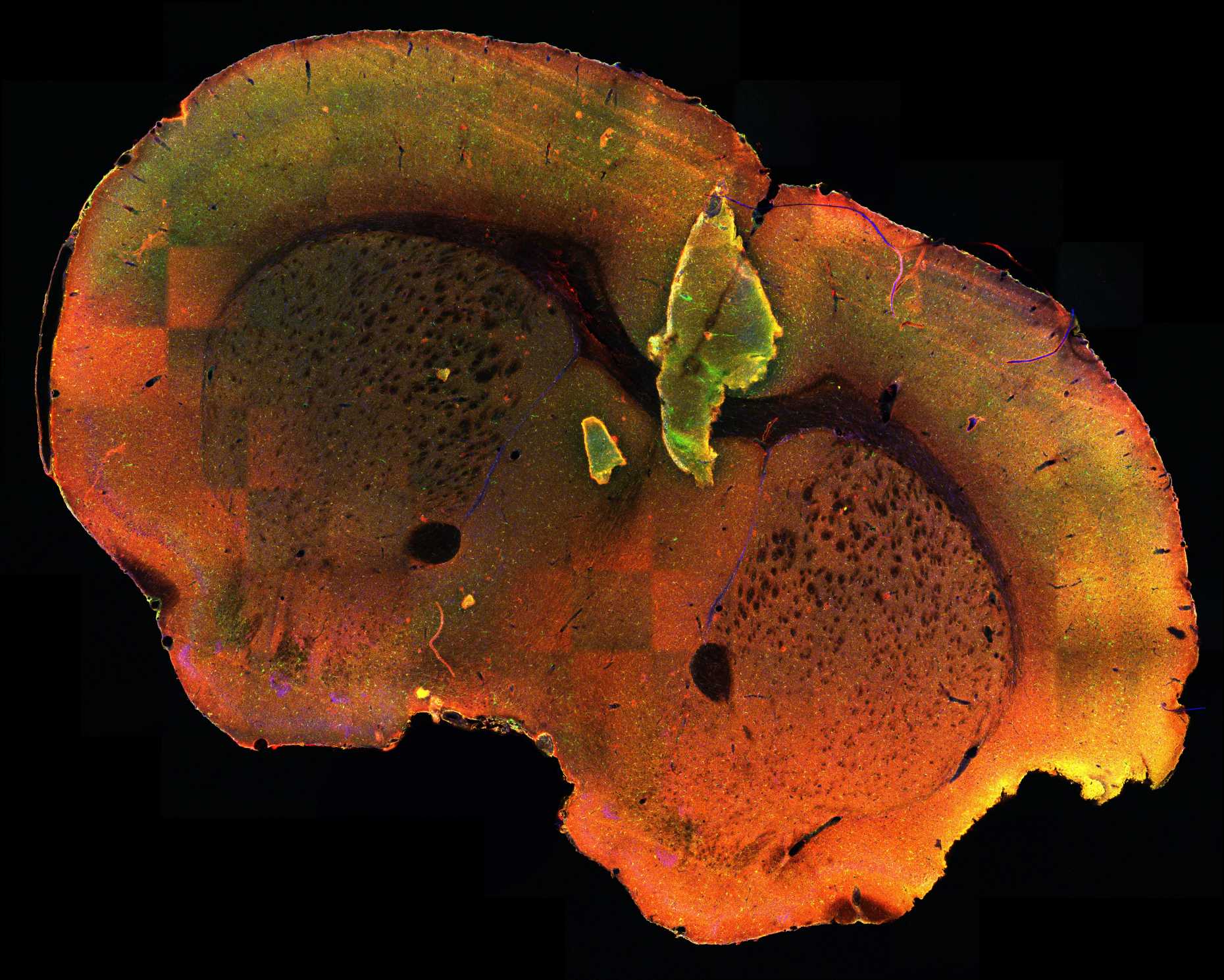
Zeiss LSM980-MP
The main features of this confocal microscope LSM980 are the nonlinear optics (NLO) and the Airyscan2 detector. The Airyscan technology alows for semi-“super-resolution” acquisition down to ~120nm. Moreover, with its so-called multiplex technology it is possible to increase the acquisition speed up to 8x while maintaining confocal resolution.
Our LSM980 is equipped with all the standard visible laser wavelengths, and additionally with two pulsed lasers (2-photon lasers, one tunable laser 680-1300 nm and one fixed frequency 1040 nm) to increase the tissue penetration capability.
The intrinsic resolution loss of 2-photon microscopy (due to the higher excitation wavelength) can be compensated for by the Airyscan detector, increasing the resolution power of 2-photon imaging.
Lastly, the system has also 2 dedicated non-descanned detectors (NDDs) for multiphoton microscopy.
Specifications:
- Multiplex acquisition (speed up acquisition to 8x)
- Airyscan detector for higher resolution
- NDD BiG module
- 32 GaAsP detector array for spectral unmixing, 2 PMT detectors + Transmitted light detector
- two 2-photon lasers for two simultaneous MP excitations with deeper tissue penetration of the excitation light
- Live cell support
- Hardware autofocus system
Full configuration details are available on the internal Wiki.
Read more about the external page Airyscan detector principle
Zeiss LSM980-IR
This microscope is a point scanning confocal that allows semi-“super-resolution” acquisition and has a dedicated laser and detectors for exciting and imaging of fluorescences also in the near-infrared range. Our LSM980-IR has 2 additional GAsP detectors and an additional 730 nm laser. With this addition you can add one extra labels/channel (emissions 760-900 nm) to your sample and/or increase the detection efficiency of the 640/Cy5 channel of your conventional confocal imaging.
The Airyscan2 detector allows imaging down to ~120nm. Moreover, with its unique multiplex technology it’s possible to increase the acquisition speed up to 8x while maintaining confocal resolution.
- Specifications:
- Multiplex acquisition (speed up acquisition by up to 8x)
- Airyscan2 detector for higher resolution
- 730nm excitation laser with 2 dedicated Infrared GaAsP detectors (660-705 nm & 760-900)
- 32 GaAsP detectrors, 2PMT detectors + Transmitted light
- Live cell environment
- Hardware autofocus system
Complete configuration information - see internal Wiki.
Read more about the external page Airyscan detector principle