Lightsheet Microscopes
The SCF houses 2 dual-side illumination lightsheet microscopes: the lightsheet 7 from Zeiss and the Viventis LS2 from Leica. These selected plane illumination microscopes (SPIM) achieve optical sectioning with a thin light sheet. This technology illuminates only the sample in the observation plane and has therefore a low phototoxicity/bleaching. Having no need for pinholes as used in confocal microscopy results also in no loss of excitation light. The signal intensity allows for short exposure times and therefore for a high temporal resolution.
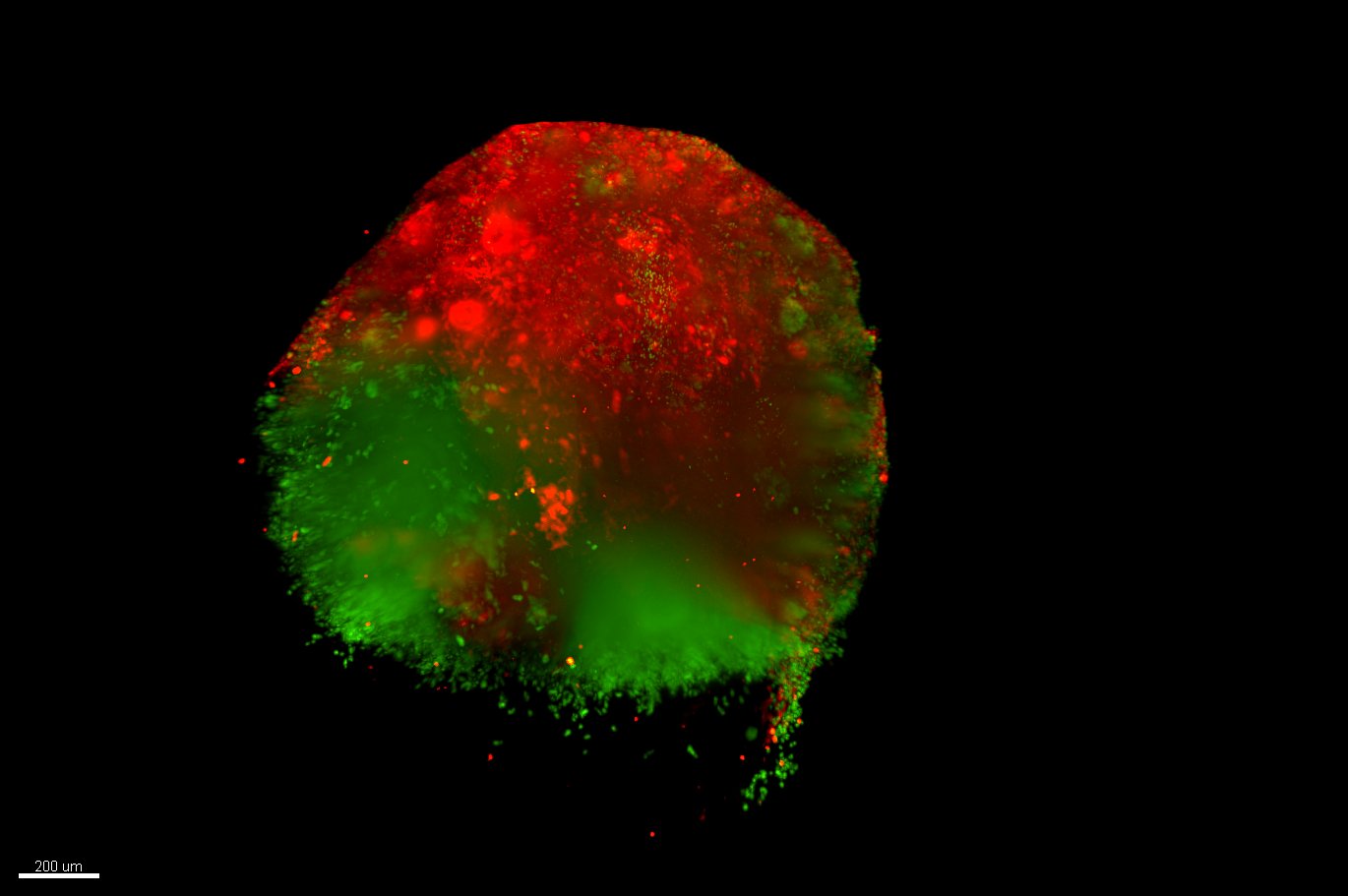
This microscopes enable users to acquire both live and fixed (cleared) samples, in a wide range of sizes and in different numbers
For detailed descriptions of the individual workstations (all specifications, filter settings, etc.) and interactive spectra viewers, see our internal Wiki (D-BSSE login required).
ZeissLightsheet 7 (Zeiss LS7) - for fixed and cleared samples
The lightsheet 7 microscope is a versatile microscope, capable of imaging fixed / cleared samples. The system is designed to mount samples vertically, using a glass capillary and hydrogel solution, and to suspend it in medium / clearing buffer. The dual-side illumination, the pivot scan that eliminates the shadows generated by any “obstacle” in the light path, and the multi-view acquisition allow to reliably obtain very uniform images. The dual camera combination enables the acquisition two fluorescence channels simultaneously, speeding up multicolor acquisitions. The system is equipped with 2 powerful workstations, one dedicated to the acquisition control with ZEN Black software, and 1 dedicated to the data collection and analysis. This configuration allows to run experiments while data are transferred to central storage or analyzed without conflicts.
Setup
- Dual illumination setup (allows better light penetration into the sample)
- Dual camera detection module: 2x PCO Edge sCMOS
- Multi-position imaging (XYZ + rotation)
- Immersion sample chamber for: aqueous media (n=1.33) or clearing media (n=1.33 - 1.58)
Excitation
- Laser lines: 405 nm, 445 nm, 488 nm, 515 nm, 561 nm, 638 nm
- Illumination optics: 5x/0.1, 10x/0.2
- Filter combination: 420-470nm + 525-545nm; 420-470nm + 575-615nm; 505-545nm + 575-615nm; 505-545nm + LP 585nm;
- Light sheet thickness: 2um - approx. 14 um
Detection
- Detection optics for water acquisitions: 5x/0.16 Dry, 10x/0.5 W, 20x/1.0 W, 40x/1.0 W
- Detection optics for cleared tissue acquisitions: 5x/0.16 Dry, 20x/1.0 Clearing (n 1.33-1.58)
- Pixel size 6.5 um
- Max image resolution 1920 x 1920 pixels
- Bit depth: 16 bits
- Max frame rate: 30 fps at 1000 x 1000 pixels
Leica Viventis Deep LS2 - for live samples
The Viventis Deep LS2 is a high-performance light sheet fluorescence microscope designed for multi-view and multi-position imaging of live samples. Its advanced design ensures optimal imaging conditions with a temperature- and CO₂-controlled sample chamber, maintaining stability even for delicate specimens.
With its patented technology, the Viventis Deep LS2 enables detailed volumetric imaging through:
• Dual illumination for enhanced clarity
• Dual-view detection for comprehensive sample visualization
• Multi-position imaging to capture multiple perspectives
• Open-top sample holder for flexible sample handling
This open-top configuration enhances throughput, allowing researchers to image multiple samples simultaneously under different conditions in a single timelapse experiment.
Ideal for various model systems—including organoids, organs as well as zebrafish embryos—the Viventis Deep LS2 ensures high-quality imaging while preserving sample viability through gentle light sheet technology.
Additionally, its advanced incubation system supports media exchange even during ongoing experiments, maintaining physiological conditions.
Setup
• Dual illumination setup (allows better light penetration into the sample)
• Dual camera detection module: Hamamatsu ORCA-Fusion
• Multi-position imaging
• Immersion sample chamber for aqueous media (n=1.33)
• Temperature and CO2-controlled
• Field of view: 25X Objective: 599µm
Excitation
• Laser lines: 405 nm, 488 nm, 561 nm, 638 nm
• Light sheet thickness: 2.3um, 4um and 7um
• two 10X objectives, NA 0.2.
Detection
• Detection optics: 25X NA 1.1, working distance 2 mm, water immersion objectives.
• Pixel size 260nm
• Field of view: 599 µm
• Available upon request two 16X Objective, FOV 936 µm;
Sample part
• Motorized XYZ sample stage with maximum X travel range of 50mm
• Multiple sample located in an open-top FEP sample chamber (multi-well).
• Sample incubation
• Recirculating air temperature controller from 5°C above ambient temperature to 40°C.
• CO2 concentration control range 2-10%.