Mapping the future of organoids
Scientists from Roche's Institute of Human Biology, their partners at ETH Zurich and Helmholtz Munich, and collaborators have built detailed atlases of brain, gut, and lung organoids to empower future discovery.
Organoids are a promising tool for better understanding disease and developing new therapies because they may more closely resemble human biology than traditional models such as animals or single cell lines in a dish. These organoids are bundles of cells made by coaxing stem cells to develop into the cell types present in organs such as the brain or intestine. However, the protocols to make organoids differ from lab to lab, and even the same protocol can generate different results in different labs. This has made it difficult to develop consistent, reproducible organoids for translational application, and compare them to the human tissues they are meant to represent.
Comparing organoids and their protocols to maximize insights
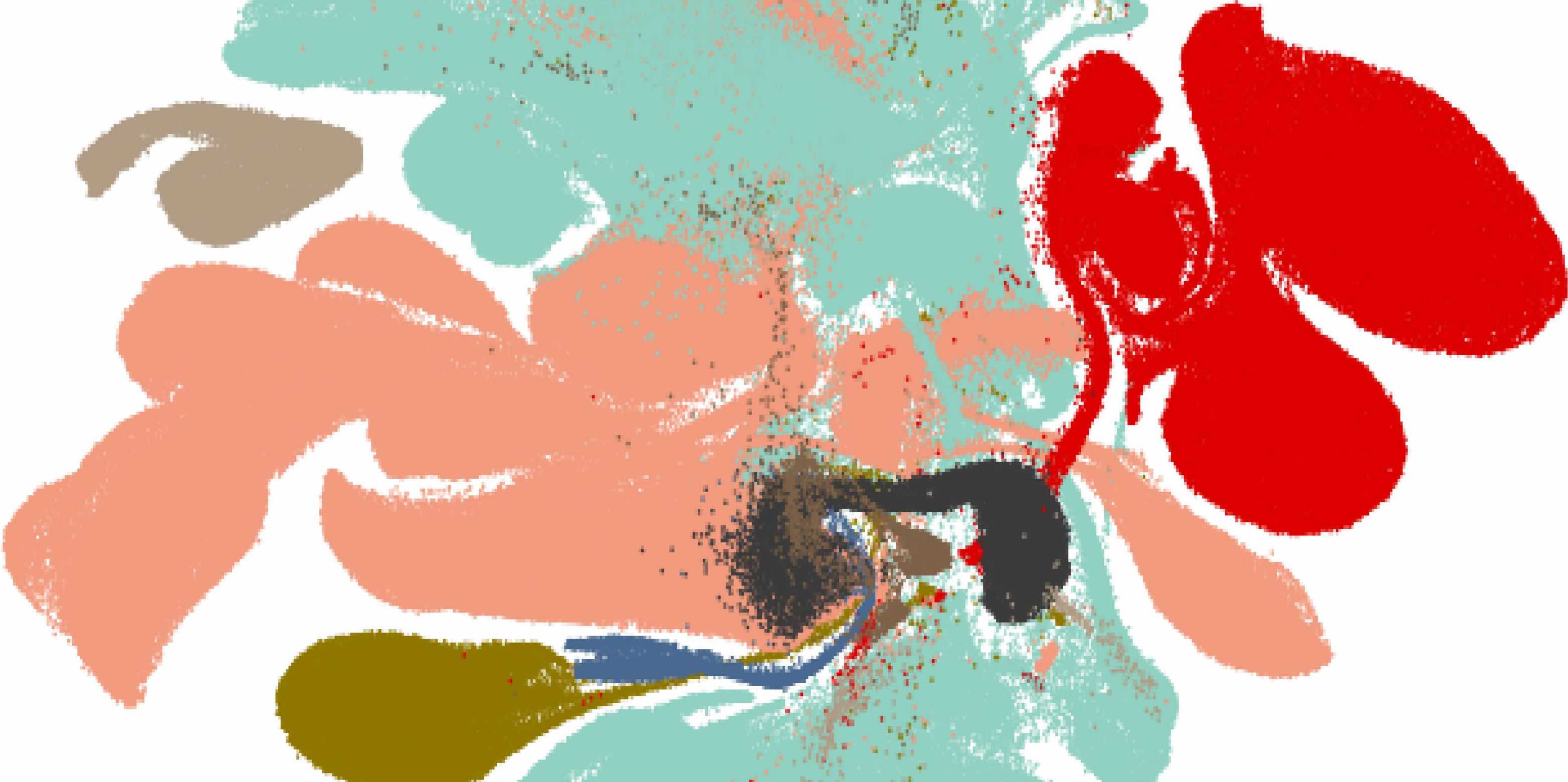
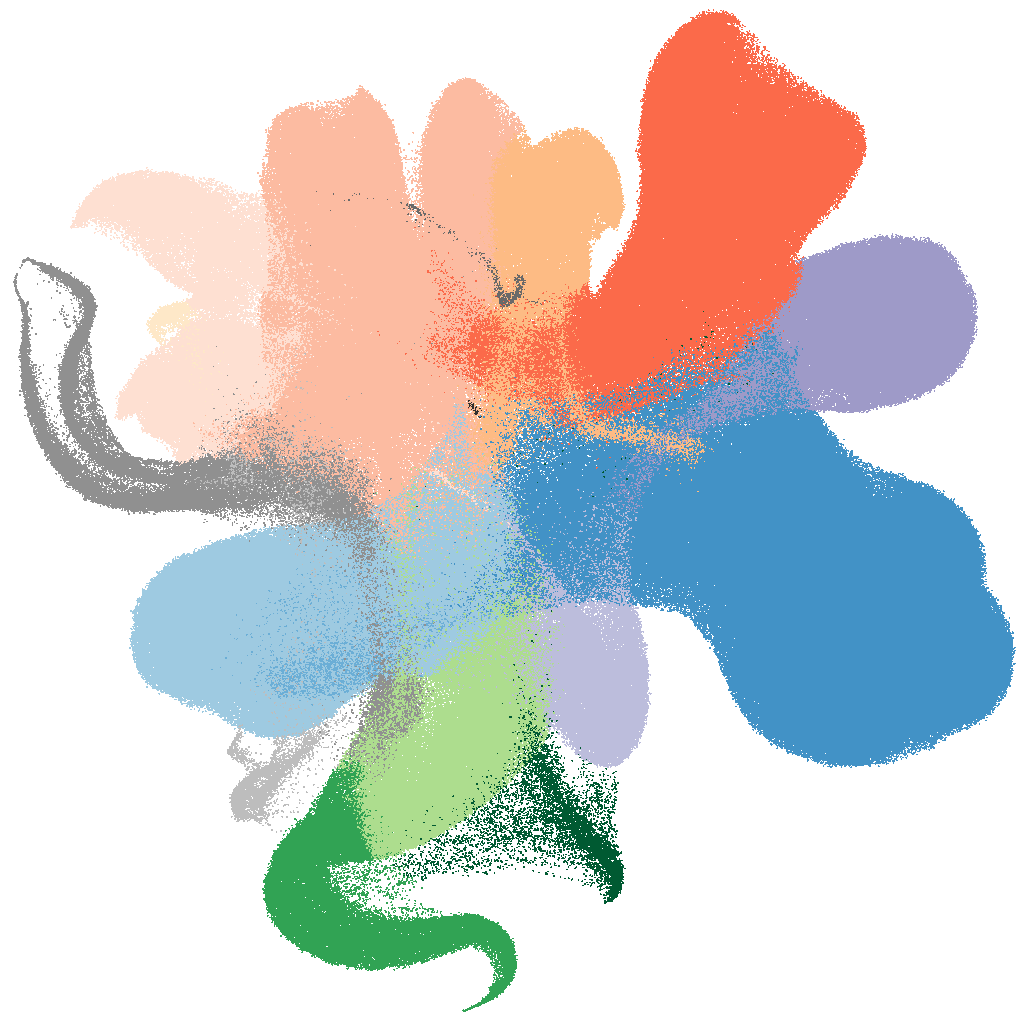
A new resource to tackle this comparison challenge at scale has now been released for organoids of both and those derived from the external page endoderm (1), and the external page brain (2) such as those mimicking the lung and intestine. Scientists from Roche’s Institute of Human Biology (IHB), ETH Zurich and Helmholtz Munich, in collaboration with academic groups across the globe have developed computational tools to integrate datasets from different protocols and labs into “organoid atlases.” These atlases now allow researchers to compare the organoids’ cells across labs, protocols, and to patient samples, including those that will be collected and studied in the future.
“For these organoid models to be used in the drug discovery pipeline, we need to understand how reproducible they are to make, what cells are present, and how we can change the models to better meet our needs,” said Gray Camp, a senior group leader at IHB and co-corresponding author on the papers.

“What we aim to get when we make organoids, and what we actually get, are not usually the same. Without the atlas, it’s more challenging to interpret the data from a single organoid protocol.”Barbara Treutlein, ETH-Professor of Quantiative Developmental Biology at D-BSSE.
Integrating diverse datasets from varied organoid protocols and labs is a difficult computational challenge, but it is essential for creating resources that are truly universal. “By harmonizing this complexity using deep representation learning, we have built atlases that not only allow us to standardize and compare organoids and the various protocols but also unlock their full potential as tools to mirror human biology,”said Professor Fabian Theis, head of the Computational Health Center at Helmholtz Munich and co-corresponding author on the papers.
Mapping first answers to key questions
Already, these atlases are answering questions that were difficult to address before. For example, the researchers showed that different protocols could generate similar cells, albeit in different proportions within the organoid. They also pinpointed cell types that were incorrectly identified before. “What we aim to get when we make organoids, and what we actually get, are not usually the same,” explained Professor Barbara Treutlein, who leads a research group at ETH Zurich’s Department of Biosystems Science and Engineering and is a co-corresponding author on the papers. “Without the atlas, it’s more challenging to interpret the data from a single organoid protocol.”
A resource of this scale also facilitates comparison to human tissue samples, helping researchers to determine what stage of human development organoids most closely resemble. Interestingly, organoids made from different starting material — such as induced pluripotent stem cells (which are reprogrammed from adult cells) or stem cells from developing or adult primary tissues — all generate slightly different stages of organoids. Without the organoid atlases, this comparison wasn’t possible.
The atlases will also be critical to enable the deployment of organoids in the drug development context. For example, this rich organoid resource can serve as a complex, diverse control for identifying new therapeutic targets. The Treutlein lab is already using the resource in this way — an unexpected benefit of generating this large, interactive data resource.
Atlases point to next steps for leveraging organoids
Moving forward, the scientists said that the atlases provide information to develop even better organoid systems, for example those that cover cell types and tissues that are currently underrepresented or that recapitulate later stages of development. As traditional organoid protocols are enhanced or replaced by new systems, such as those external page incorporating immune cells or into external page organs on a chip, these resources will provide helpful guides to develop the most useful and informative models.
In addition to being a collaboration between the IHB and academic researchers worldwide, the organoid atlases form part of the international Human Cell Atlas (HCA) consortium, which is creating comprehensive reference maps of all human cells as a basis for both understanding human health and diagnosing, monitoring, and treating disease. Camp noted that the HCA will be a critical resource to ensure that organoids do, in fact, represent human biology. “It’s an important emerging field to be building accurate human model systems to test therapies. The more people who can be involved, the better,” he said.
“We want these atlases to have an impact, so we have to have everyone on board,” agreed Treutlein. “This was a fantastic and productive collaboration with many of the world leaders in in vitro systems.”
The organoid atlases presented in the papers are just the beginning. Looking ahead, the integration of organoid models with cutting-edge AI tools will drive unprecedented opportunities to study human biology at scale. “Fine-tuning the used protocols to individual needs via generative AI will bring us closer to personalized medicine, enabling the creation of organoids tailored to individual patients and specific therapeutic needs,” said Theis. This convergence of biology and computation is paving the way for the next generation of human disease modeling and drug discovery.
About Human Cell Atlas (HCA): The Human Cell Atlas (HCA) is an international collaborative consortium whose mission is to create comprehensive reference maps of all human cells—the fundamental units of life—as a basis for understanding human health and for diagnosing, monitoring, and treating disease. The papers mentioned in this article are part of a collection of more than 40 HCA publications in Nature Portfolio journals that represent a milestone leap in our understanding of the human body. As an open, scientist-led consortium, HCA is a collaborative effort of researchers, institutes, and funders worldwide, and will provide a foundation to transform and democratise global healthcare. external page https://www.humancellatlas.org/
Find the original News article and additional resources on the external page Roche corporate website.
Journal papers are available open access
- Xu Q*^, Halle L*, Hediyeh-zadeh S, Kuijs M, Riedweg R, Recaldin T, Yu Q, Rall I, Frum T, Adam L, Parikh S, Kfuri-Rubens R,Gander M, Klein D, Curion F, He Z, Fleck JS, Oost K, Kahnwald M, Barbiero S, Mitrofanova O, Maciag GJ, Jensen KB, Lutolf M, Liberali P, Spence JR, Gjorevski N, Beumer J, Treutlein B^, Theis FJ^, Camp JG^ (2025). external page An integrated transcriptomic cell atlas of human endoderm-derived organoids. Nature Genetics. https://doi.org/10.1038/s41588-025-021826
*lead authors
^senior corresponding authors He Z*^, Dony L*, Fleck JS*, Szałata A, Li KX, Slišković I, Lin HC, Santel M, Atamian A, Quadrato G, Sun J, Pașca SP, Human Cell Atlas Organoid Biological Network, Camp JG^, Theis FJ^, Treutlein B^ (2024) external page An integrated transcriptomic cell atlas of human neural organoids. Nature doi.org/10.1038/s41586-024-08172-8
*lead authors
^senior corresponding authors