Mapping the axolotl forebrain and its regeneration
Axolotl salamanders are amphibians that have the extraordinary ability to regenerate brain tissue after injury, yet their brain is largely unexplored. In a study published in Science, an international team of researchers from Barbara Treutlein’s lab, in collaboration with Elly Tanaka’s lab at the Institute of Molecular Pathology in Vienna, Austria, map out all cell types of the axolotl forebrain, and study the formation of new neurons during regeneration.
The forebrain has been under intense selective pressure throughout evolution because it plays a key role in animal behaviour and cognition. Single-cell and single-nucleus sequencing technologies can be used to shed light on the cellular composition of the brain as it allows researchers to measure which genes are active in individual cells providing a way to group cells into different cell types and to understand their function. These technologies have been applied to the brains of fish, reptiles, and mammals, yet the cellular composition of the amphibian brain and hence its functional organisation has so far remained a mystery.
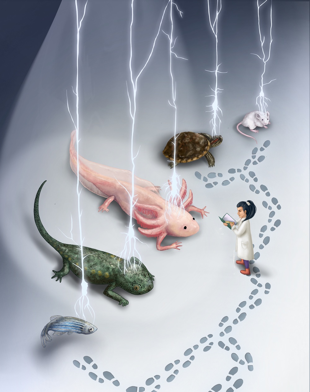
Now, in a study published in Science, Ashley Maynard, Tomás Gomes and other researchers from the Quantitative Developmental Biology lab led by Barbara Treutlein, in collaboration with researchers from Elly Tanaka’s lab at the Institute of Molecular Pathology in Vienna, Austria, used single-nucleus sequencing to characterise with unprecedented detail the cell populations of the axolotl forebrain. The atlas revealed close to 100 cell populations including neuronal and non-neuronal cell populations. The team then compared the identified cell populations with cell atlases of turtle and mouse brain, which have been extensively studied. This revealed that the axolotl brain contains neurons that correspond to the mammalian hippocampus, the lateral cortex, and potentially even to the cerebral cortex.
Axolotl brains have life-long neurogenic activity, meaning new neurons are continuously made throughout the life of the axolotl. For the first time, this study resolved the paths from neural progenitor cells through intermediate stages to neurons in the different parts of the axolotl forebrain. In addition, the authors used a external page computational strategy recently developed in the Treutlein lab to illuminate which gene networks are controlling the differentiation from the progenitor to the neuron state.
Axolotls are famous for their prodigious capacity to regenerate after injury - including complete removal of large parts of the forebrain. The authors used a modified single-cell sequencing strategy to explore what happens in response to injury and uncovered that progenitor cells rapidly multiply and a subset of them activates a regeneration-specific wound-healing state. The cells then differentiate to restore all neuronal populations that have existed before the injury and neurons even restore their connections to other brain regions. This indicates that the regenerated brain regains its functionality.
This cell atlas from the axolotl forebrain has exciting implications for translational medicine: Refurbishing the regeneration potential of neuronal cells in humans would step up regenerative medicine and provide new alleys to treat neurodegenerative diseases.
Find original publication:
Lust*, K, A Maynard*, T Gomes*, J S Fleck, J G Camp, E M Tanaka, and B Treutlein (2022) external page Single-cell analyses of axolotl telencephalon organization, neurogenesis, and regeneration. Science, doi 10.1126/science.abp9262.
*These authors contributed equally to this study.
Find external page Science Press Package on EurekAlert.
external page Media release issued by the Institute of Molecular Pathology, Vienna, Austria.
Learn about the Quantitative Developmental Biology lab led by Barbara Treutlein.