Studying the mechanosensitivity of neurons at nano-scale
Right from the early developmental stages to fully grown adults, brain cells continuously rely on mechanical cues to migrate to the correct locations in the body, form connections with other neurons or other cell types and communicate with them in a unique language of electrical impulses. In a new study, researchers from the Biophysics group of Daniel Müller and the Bio Engineering Lab of Andreas Hierlemann reveal how neurons sense the magnitude and temporal features of physical forces.
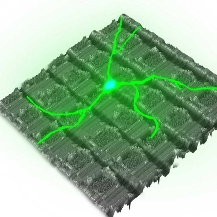
Studying the brain, a mechanosensitive organ, requires precise tools that can see neuronal cells in action, catch the forces at play, and listen in on their electrical conversations, in other words: tools that mechanically stimulate and simultaneously record the electrophysiological response of neurons within neuronal networks. By combining atomic force microscopy (AFM), fluorescence microscopy and high-density micro-electrode arrays, the group of researchers led by Krishna Kasuba measures and characterises the mechanical properties of neurons at nano-scales. One key finding: Neurons differentiate between various mechanical stimuli and functionally respond in a different manner. This line-up of tools opens the door to explore new ways of mechanical characterisation, stimulation and control of complex electrogenic biological systems in organoids and tissues.
Find research article in Nature Nanotechnology:
Kasuba, K C, A P Buccino, J Bartram, B M Gaub, F J Fauser, S Ronchi, S S Kumar, S Geissler, M M Nava, A Hierlemann and D J Müller (2024) external page Mechanical stimulation and electrophysiological monitoring at subcellular resolution reveals differential mechanosensation of neurons within networks. Nature Nanotechnology, https://doi.org/10.1038/s41565-024-01609-1
Learn about the Biophysics lab led by Daniel Müller, the Bio Engineering Lab of Andreas Hierlemann.